Sensors Provide a Clear Vision for Minimally Invasive Surgery
Sensor-guided technologies help surgeons push the barriers of surgical innovation.

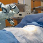
Thanks to sensor-guided medical technologies such as robot-assisted surgery, endoscopic diagnostics, and electromagnetic navigation bronchoscopy, surgeons are able to perform precise medical procedures with minimally invasive surgery (MIS), accelerating patient recovery and improving outcomes. Robotic arms combined with 3D visualization of the site allow doctors to access internal structures through a tiny incision and perform microscopic procedures with precision and control. Force, position, and torque sensors guide the instruments. Temperature sensors help monitor the patient during anesthesia.
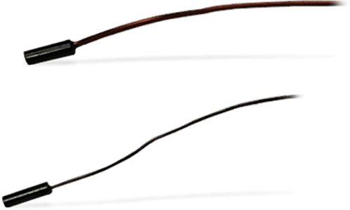
Amphenol Advanced Sensors Type SC Series, available from Mouser Electronics, includes miniature chip-in-glass or sleeved chip thermistors with fine diameter wires for insertion into hypodermic needles for myocardial surgeries and external attachment to metal lumens used during laser surgery.
Another area of medicine where sensors help deliver care is endoscopy. Endoscopic instruments mounted on flexible tubes are typically used with video monitoring for ear, nose, and throat procedures. They are also used for electrophysiology procedures for heart patients and complex operations on the lungs, the spine, and in neurosurgery. Endoscopic cameras use sensors in combination with optical interconnects to create enhanced images of internal body structures.
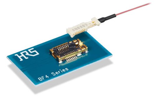
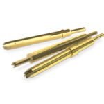
Hirose Electric’s extensive selection of connectors for endoscopes includes the BF4M Series Optical Active Connector, which delivers high-speed signal transmissions with no EMI noise.
In electromagnetic (EM) navigation systems, a signal tracks the instrument within the patient to reach a growth. An example would be an endoscope negotiating the myriad branches of a lung. The electromagnetic field guides the instrument to the area of interest, providing access to areas which were previously inaccessible to traditional catheter techniques. These surgical navigation systems are used in operating rooms as well as outpatient centers and walk-in surgical centers.
Electromagnetic fields capture and relay sensor position, overlaid on the patient’s anatomy and organs, so that the surgeon can establish the precise location of the surgical instruments. Growths and tumors in the brain, for example, can be hard to accurately locate and reach. Precise location of the surgical tool minimizes contact with healthy tissue and reduces the risk of damage to blood vessels and nerve endings in the process.
Metallic objects, or multiplex magnetic generators and receiver coils – known as localizers and trackers, respectively – are used to stabilize the signal. An accurate signal improves surgical precision, despite the surgeon not having access to a clear line of sight.
EMI in the OR
Surgical tables and patient beds often contain conductive material that can cause distortions through electromagnetic interference (EMI). They are static structures, however, so the interference from them can be mapped before the procedure and the data used to correct the patient-instrument location during surgery.

“In today’s MIS and robotic procedures, there are a lot more imaging equipment and robotic arms in the procedure room; their presence speaks to EMI. In spinal surgery, for example, there are a lot of metal structures in the procedure space: the fluoroscopic C-Arm, metal robot part, along with various surgical and fixation tools. Since these move around, in and out of the surgical field, they can’t be mapped prior to the procedure. EM systems must detect when there is EM interference present and also mitigate the location distortion caused by them,” said Andrew Brown, CEO of Radwave, a company that has developed electromagnetic tracking solutions for medical devices.
Conventional bulky EM field generators are incompatible with commonly used imaging equipment for procedures like CT (computerized tomography) or fluoroscopy. They also have limited or no detection or correction of EM-induced distortions, leading to inaccuracies, explained Brown. “This is especially true in environments where additional equipment such as fluoroscopes, robots, and other metal objects are often used.”
Frequency interference is also an issue, he said. Some older EM systems use wide electromagnetic frequency bands which often create interference with medical equipment like an ECG (electrocardiogram) for example, or an image from a fluoroscopic C-Arm. This means the older EM systems cannot be used simultaneously or in proximity to each other.
The company has developed a customizable, modular electromagnetic tracking platform for diagnostic and therapeutic devices used during surgical procedures. It consists of a control unit and a polycarbonate antenna (or field generator) which is placed between the patient and the operating table or can be integrated into the table. The third element, developed in collaboration with TT Electronics, is a selection of sensors with navigation options of five or six degrees of freedom (DOF).
When the device is paired with an EM tracking system, the sensor shows the physician the precise location of the device during the procedure. “Regarding the sensor geometry, different sensor geometries and shapes, sensor design configurations, and number of sensors within the assembly can drive the 5DOF or 6DOF navigation characteristics of the device,” said Garrett Plank, business development director at TT Electronics.
Successful surgeries depend on the skilled hands and sharp eyes of the surgical team. But as that team continues to expand to include technology partners, the use of a variety of miniaturized sensors will continue to populate the operating theater.
To learn more about the companies mentioned in this article, visit the Preferred Supplier pages for Amphenol Communications Solutions and Hirose Electric.
Subscribe to our weekly e-newsletters, follow us on LinkedIn, Twitter, and Facebook, and check out our eBook archives for more applicable, expert-informed connectivity content.
- Matter: A Show of Unity for Connectivity - September 5, 2023
- Brexit Update: UK Connector Industry - March 28, 2023
- Sensors Make it Plain Sailing for a Smart Ferry - February 21, 2023
