Top Trends in the Diagnostic Imaging Equipment Market
Imaging equipment is evolving rapidly based on technological advancements, miniaturization, and portability. Connectivity solutions are scaling down in size and up in capability to enable these advanced designs.
Diagnostic imaging equipment enables physicians to obtain digital images of structures inside the body in a non-invasive procedure. These images can identify potential abnormalities behind skin, tissue, and bones to guide treatment decisions.
Technology is rapidly changing the capabilities, dimensions, and flexibility offered by imaging equipment. The big iron imaging devices of the past are becoming increasingly smaller and more portable. Magnetic resonance imaging (MRI) produces 3D anatomical images by detecting the change in the direction of the rotational axis of protons found in the water inside living body tissues. An MRI scanner applies a very strong magnetic field (0.2 to 3 tesla) — about 1,000 times greater than a typical refrigerator magnet — which aligns the magnetic field and radio waves as the proton “spins” to generate images of the body that cannot be seen as well with X-rays, computerized tomography (CT) scans, or ultrasound. These MRI systems primarily use small, multi-function non-magnetic connectors and interconnect cable harnesses.
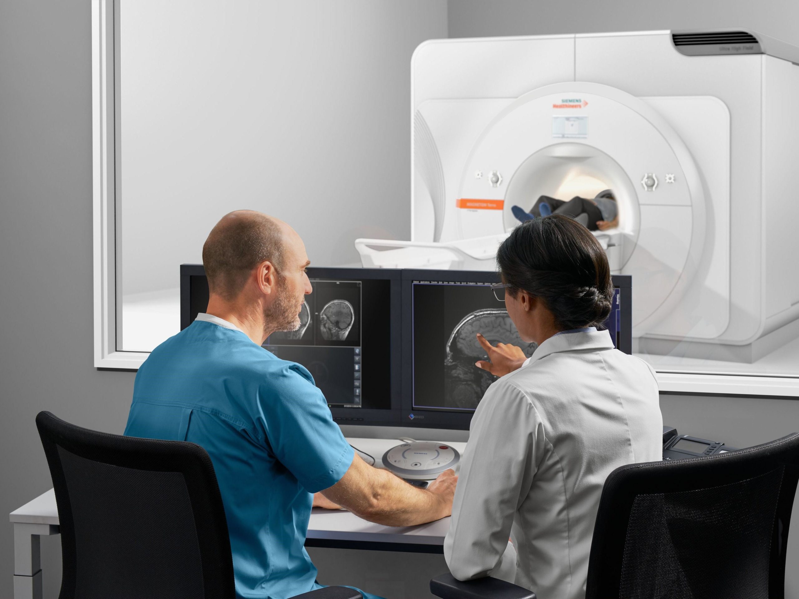
Siemens Healthineers 7T Magnetom Terra MRI System more than doubles the static magnetic field strength. This advanced ultra-high-field (UHF) technology is primarily used for neurological and musculoskeletal examination of the head, arms, and legs.

Open MRI machines, such as the Hitachi APERTO Lucent Open MRI System, offer patients a more calming experience while powerful gradients deliver fast and reliable high-resolution diagnostic images.
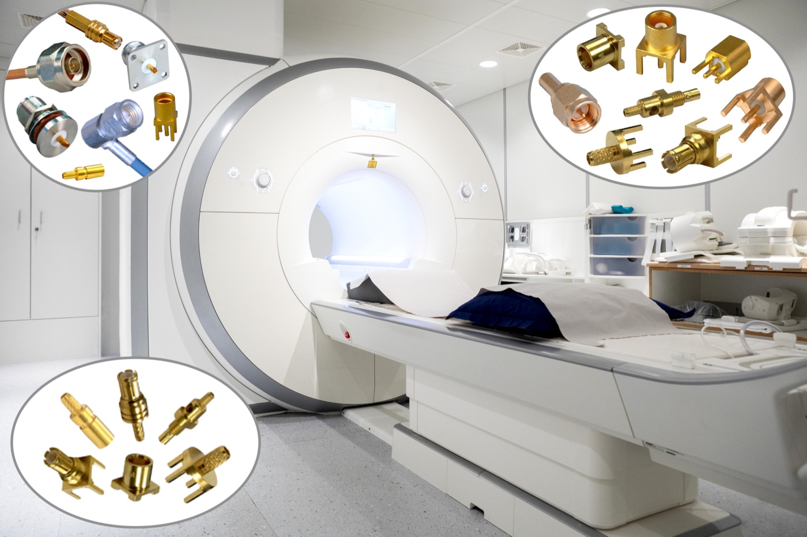
Cinch Connectivity Solutions offers an expanded range of non-magnetic connectors with new SMA, SMB, and MCX interface versions. These connectors are well-suited for MRI and magnetic resonance angiography (MRA) equipment.
Non-magnetic RF Connectors are essential components in advanced MRI systems. Connector suppliers that offer these products include AirBorn, Amphenol Pcd, Fischer Connectors, Molex, Nicomatic, Radiall, Rosenberger, SV Microwave, Samtec, Smiths Interconnect, and others.
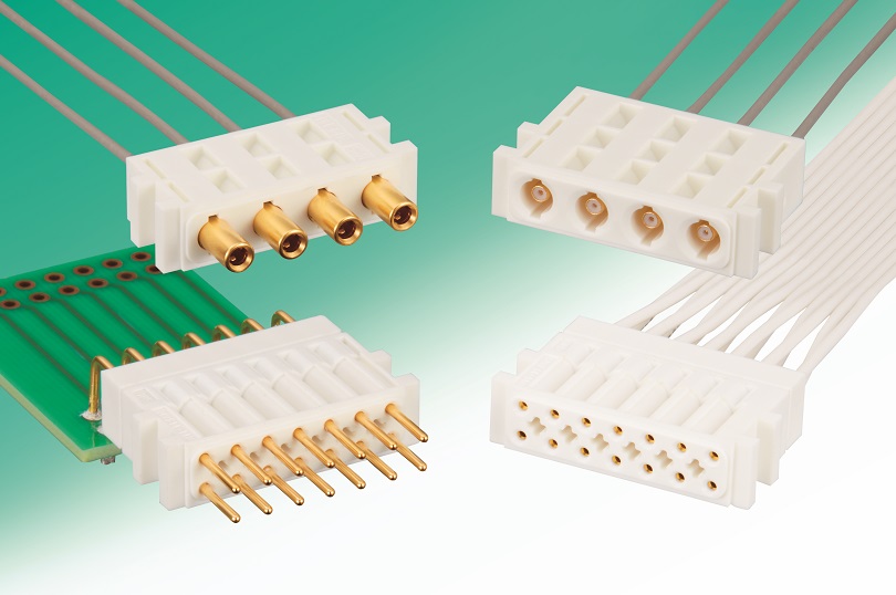
Hirose MRF14 Series coaxial high-durability connectors are non-magnetic, rated for more than 60,000 mating cycles, have a low insertion and extraction force, come in various customizable housing designs, and are available as solder type for cable termination.
Siemens Healthineers is one of several medical technology companies that have developed mobile MRI systems that are housed within tractor trailer trucks. These units move from place to place to treat patients who don’t have access to clinical imaging services, such as those located in remote, rural, or underserved areas. In addition to the typical MRI connectors and cables needed for a fixed MRI installation, mobile MRI stations need robust, modular, high-voltage connectors and cable assemblies to bring power from its source to portable MRI trailers.
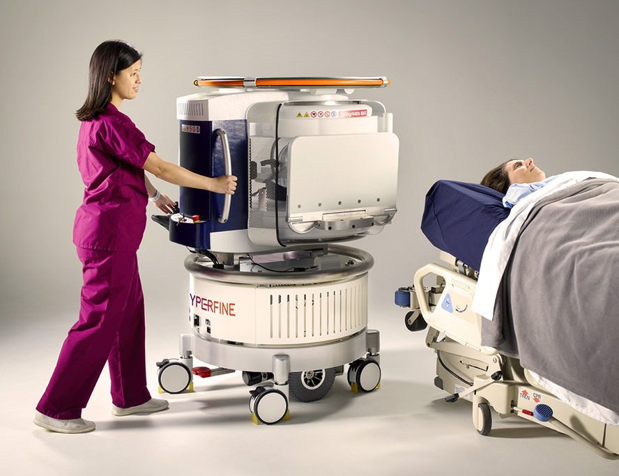
Smaller portable MRI systems are emerging to bring imaging diagnosis closer to the patient. In 2020, the FDA approved the Hyperfine Swoop, the world’s first portable MRI. This device can be wheeled to a hospital bedside for neuroimaging of a patient’s head and brain. Hyperfine plans to extend diagnosis to the knee, foot, hand, wrist, elbow, shoulder, spine, and other regions of the body. This portable imaging system weighs about 90% less than standard stationary MRI systems and fits in elevators. It runs off conventional power and renders clinical high-resolution 3D images.
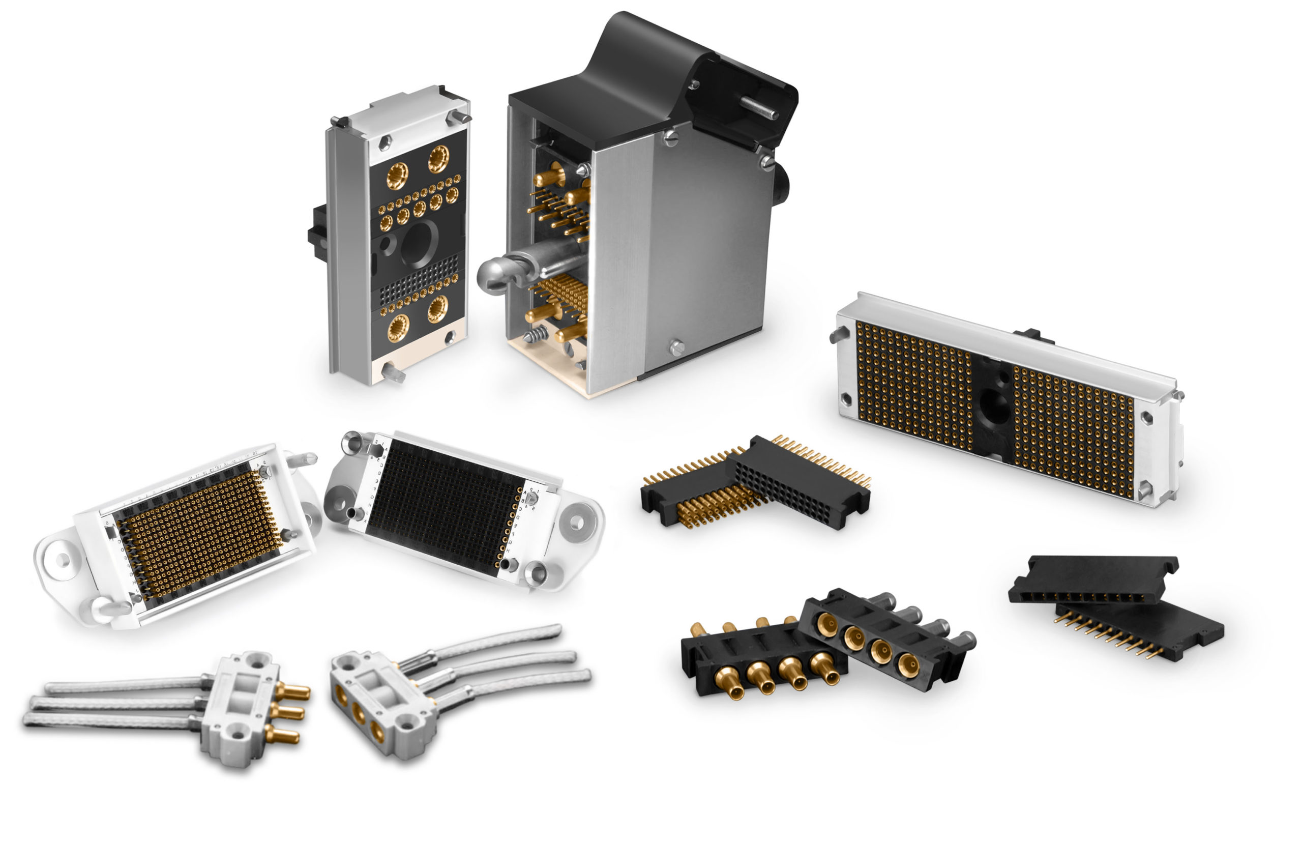
Smiths Interconnect’s N Series of high-density mini-modular connectors use the building block principle to package high-density contacts in a modular system that enables a variety of combinations in a single connector frame. Designers can create a custom connector by selecting the components that fulfills the specific electrical, mechanical, and environmental performance requirements with off-the-shelf components.
Technological Advancements Expand Market Adoption
Unlike MRI systems, CT scanners use computerized X-ray imaging to generate cross-sectional image “slices” of the body that can be digitally stacked together to form 3D images of bones, blood vessels, and organs. These images can detect tumors or other abnormalities. The emergence of low-dose and automated CT scanners offers high image quality with improved spatial resolution and low radiation exposure to the patient and healthcare professionals. Advancements in semiconductors have enabled device miniaturization and the growing trend toward portability. Technically advanced cone beam computed tomography (CBCT) systems are expanding the applications in which modern CT imaging equipment can assist. These developments are contributing to increased global market adoption.
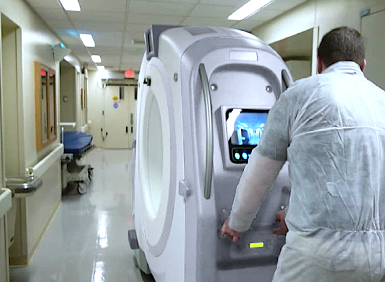
Neorologica, a subsidiary of Samsung, introduced a portable, full-body, 32-slice, CT scanner called the BODYTOM. The system has an 85cm gantry and 60cm field of view, the largest field of view available in a portable CT scanner. The BODYTOM is a battery-powered portable CT designed to accommodate patients of all sizes in clinical settings or in specially equipped vehicles for community-based care.
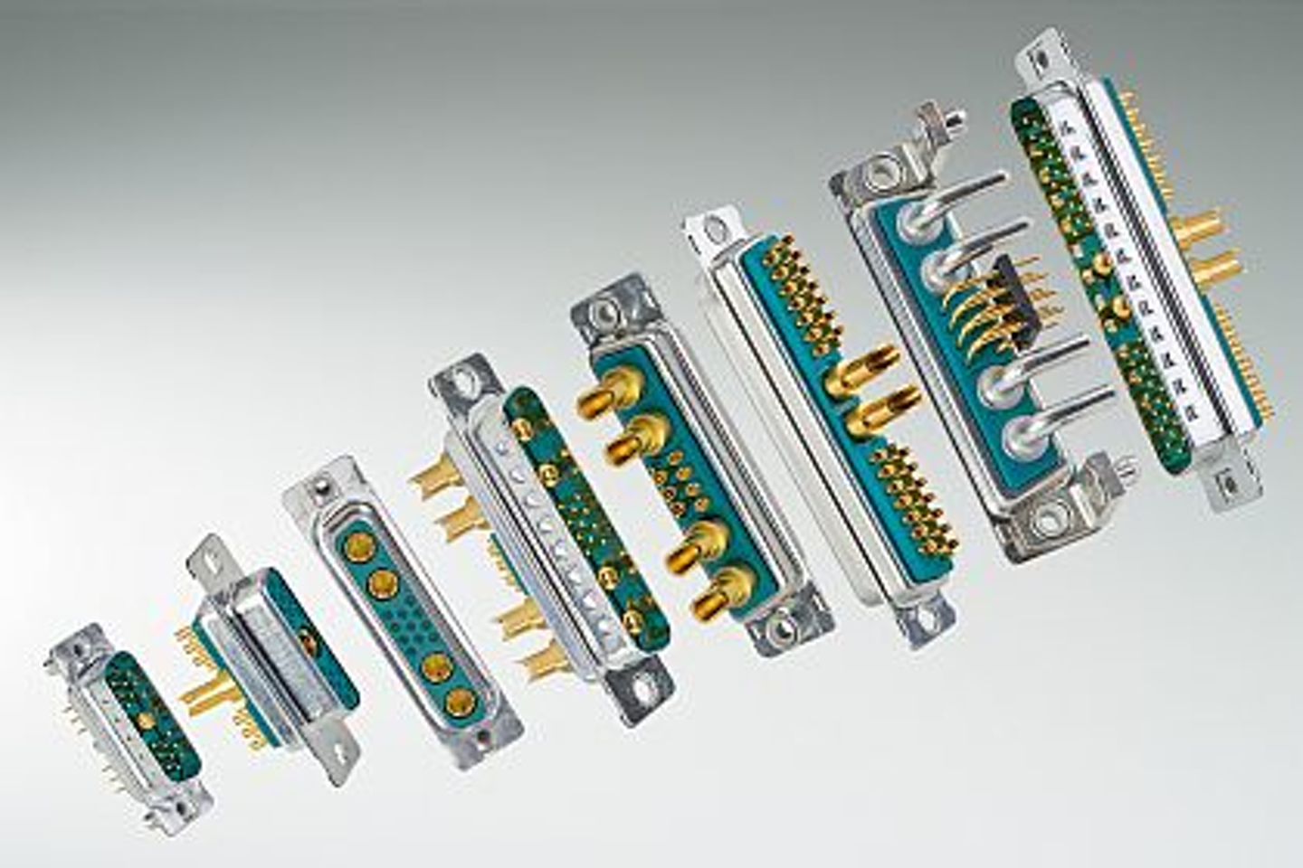
CONEC offers a series of non-magnetic combination D-subminiature connectors that are suitable for use in advanced medical imaging equipment. Combo D-sub connectors provide a high signal-to-noise ratio (SNR).
Comprised of copper alloy shells, tin-plated brass components like screws or threaded rivets, and diecast zinc brackets, the non-magnetic combination D-sub connectors can include signal, high power, or coaxial contacts. The advanced non-magnetic combination D-subs are available in 21 standard pin configurations, as well as in custom arrangements. CONEC non-magnetic D-subs combine small size and light weight characteristics with high performance and ruggedness.
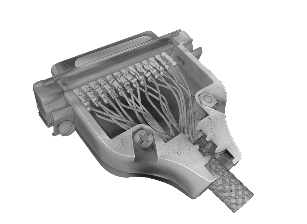 North Star Imaging, an ITW company, uses CT equipment to evaluate electrical connector designs with multiple pins and complex geometries. The imaging technology is used to inspect for wire damage, contact-to-wire insertion positions, pin positions, foreign objects, debris, insulation integrity, and position. In addition, connector shells and overmolds can be inspected for shrinkage, cracks, porosity, and other casting discontinuities. While NSI offers this as a service, many connector and cable assembly suppliers are investing in CT equipment to diagnose wiring and molding abnormalities, as well as for validation purposes.
Miniaturization Fuels Portability
Diagnostic ultrasound imaging systems are evolving from portable carts to laptop and tablet-based systems to handheld and wireless systems. Portability trends are driving higher contact density and miniaturization in the connectors used inside these systems. Portable ultrasound cart systems have numerous zero insertion force (ZIF) receptacles designed to support four, six, or even more transducers. Ultrasound connectors and cables are typically supplied in sub-assembly form. The product consists of a ZIF connector that contains complex electromechanical content, housed in a durable shell casing, terminated at the distal end to transition boards or with FPC style connectors.
Charles Staley, global industry marketing manager for I-PEX, said, “High-resolution imaging requires faster signal speeds and larger data handling and memory processing capacity without compromising signal integrity. To accomplish this objective, I-PEX micro-coaxial assemblies replace traditional flex circuit transition boards with 42AWG micro-coax and its CABLINE connectors that are lower loss and less susceptible to electromagnetic interference (EMI), thus improving performance at high speeds.”
I-PEX uses a robotic feed system to create micro-coax cable bundles, and proprietary robotic assembly lines to cut, strip, and prep fine AWG micro-coax for highly repeatable and consistent lead-frame solder processing, said Staley. “A leading CT imaging equipment manufacturer recently selected I-PEX for a wire harness combining CABLINE-UM connectors with micro-coax signal and power lines for its state-of-the-art CT machine that required 2.5GHz signaling with low profile and high-density termination.”
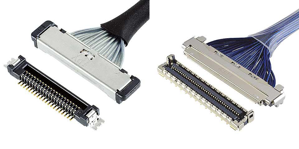
The I-PEX CABLINE® -UM is a 0.4 mm pitch, vertical right angle micro-coaxial wire-to-board connector with 360° EMI shielding, multi-point ground design for use with 36 to 46AWG micro coax. The connector can accommodate 30- and 40-pin configurations.
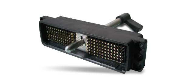
ITT Cannon’s DL Series ZIF Connectors have a CAM actuation lever for mating up to 360 high-density contacts with multiple-wire power and signal connectors. DL connectors are rated for a minimum of 10,000 mating cycles without performance loss.
The handheld ultrasound market is growing rapidly, as it gains acceptance among physicians due to its flexibility and cost advantages. Global sales of handheld ultrasound equipment are expected to increase by over 50%, to exceed $500 million by 2023.
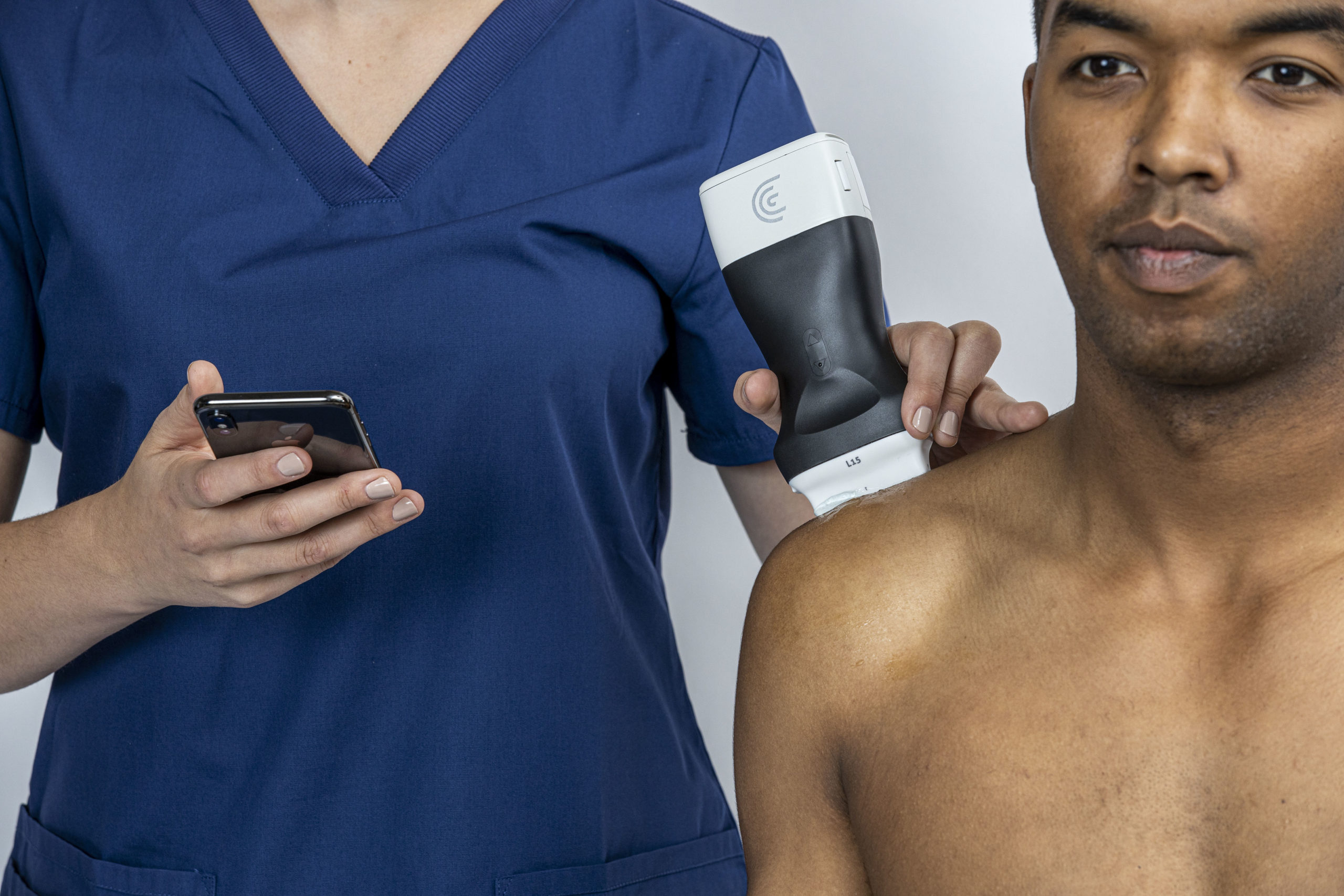
Clarius has designed wireless hand-held ultrasound units to bring portable, cost-effective imaging technology to more patients.
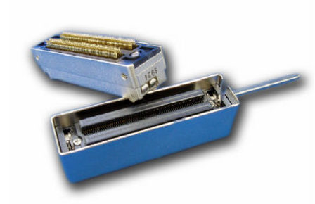
The ITT Cannon Miniature QLC connector is designed for use in portable medical ultrasound equipment. The Canon QLC is 60% smaller than the Canon DL connector with the same number (260) of contacts. The mating receptacle is available in standard solder tail and press-fit contacts, enabling easier termination. The QLC is rated for a minimum of 20,000 mating cycles, whereas the DL is rated for 10,000 cycles. The interface utilizes EMI springs and shield-locking mechanism to ensure an even mating pressure around the perimeter of the mated connector, creating an EMI/RFI shield.
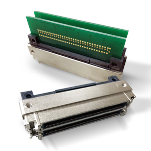
The ITT Canon QLC solderless plug is a 260-pin ZIF connector that eliminates the need to hand solder fine-pitch contacts onto transition boards, thus reducing assembly time and labor costs. The QLC solderless plug is fully compatible with existing receptacles.

